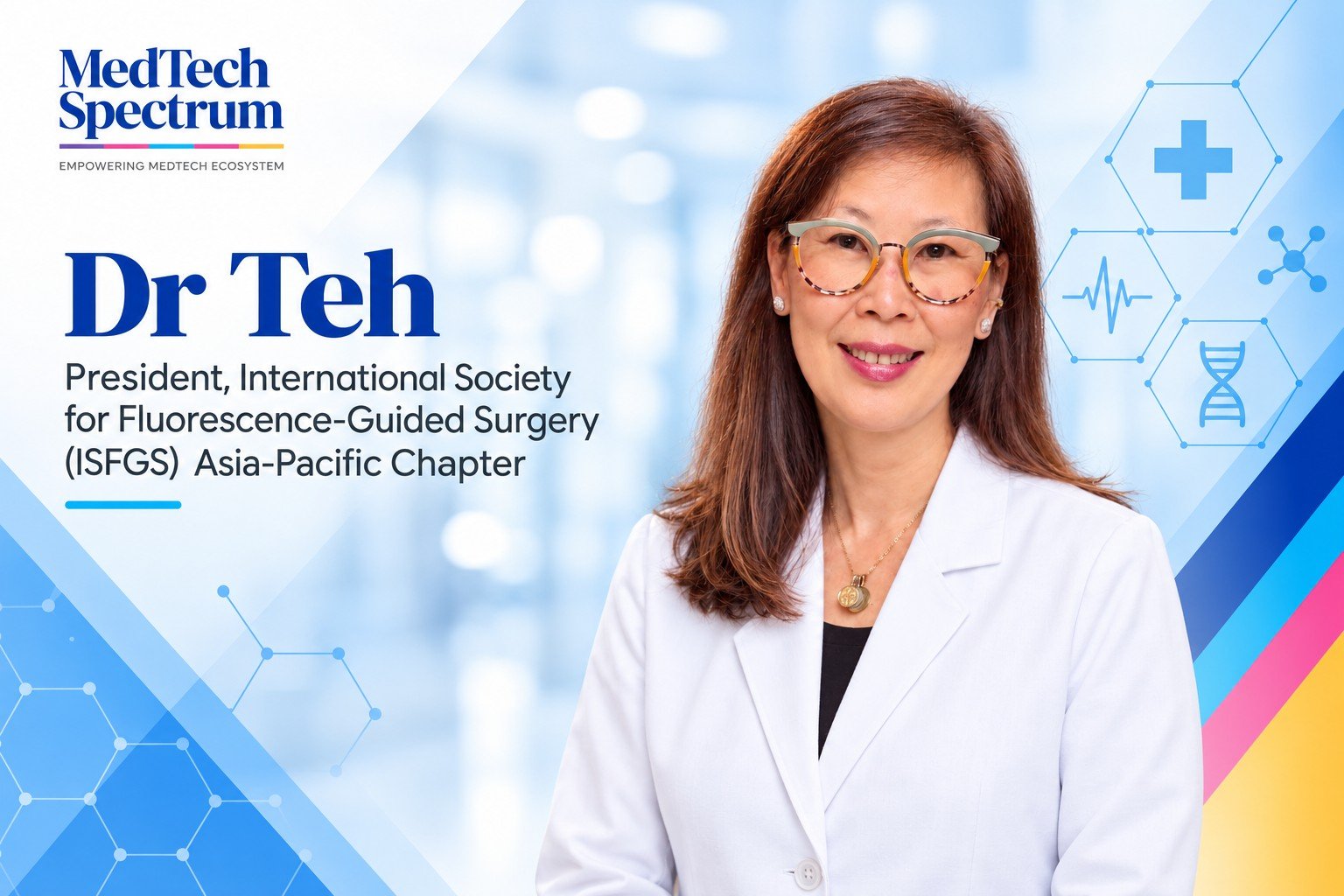
Robotic platforms, intraoperative ultrasound and fluorescence imaging are expanding the information and precision available to surgeons. In complex liver procedures, however, their value depends on how clinicians interpret those signals, reconcile conflicting findings and adapt when the operative reality departs from the original plan.
For Dr Catherine Teh, a hepatopancreatobiliary surgeon, a recent robotic segment VIII liver resection illustrates this relationship. Combining ultrasound and indocyanine green (ICG) fluorescence with a nasogastric tube adapted for flexible suction, the procedure highlights how advanced technology and practical clinician-led innovation can work together in a constrained operative field.
In this interview, Dr Teh discusses the limits of automation, the evolving role of the surgeon as a clinical integrator, and why validation, training and equitable access will shape the impact of image-guided surgery across Asia.
Robotic surgery continues to advance rapidly, but your recent case highlights the importance of clinician judgement and adaptability. In what situations does human expertise remain critical despite increasingly sophisticated surgical technologies?
Human expertise becomes most important when the operative reality departs from the plan. A robotic platform can provide magnified three-dimensional vision, stable instrumentation and fine control, but it does not understand the significance of unexpected anatomy, poor tissue quality, evolving haemorrhage or a change in the patient's physiology. The surgeon must integrate all of those signals, decide which information to trust, and know when to modify the technique, convert the approach or stop.
One of our recent segment VIII liver resections illustrates that point. The hepatic dome is technically demanding because access is constrained, visual angles are limited and conventional rigid suction can compete with the working instruments. Robotics gave us precision, while intraoperative ultrasound defined the tumour's relationship to the major vascular structures and ICG fluorescence helped with segmental demarcation and bile-leak assessment. We then adapted a standard nasogastric tube as a flexible suction device to maintain exposure in a difficult space.
That adaptation was not separate from the technology; it was what allowed the technology to work in the patient's actual anatomy. In complex surgery, judgement and decisive innovation or adaptation are part of a continuous process that determines how, when and whether each tool should be used.
Fluorescence-guided surgery is gaining traction across multiple specialities. How is this technology improving intraoperative decision-making, and what benefits are you seeing in complex procedures?
ICG in liver surgery is an evolution, not a completely new concept. The dye entered clinical use in the 1950s and has been used for decades to assess hepatic clearance. In Asia, ICG-R15 became deeply embedded in preoperative liver assessment, including the Makuuchi decision criteria for the safe extent of hepatectomy. The fluorescence era came later: in the late 2000s, Japanese groups demonstrated ICG-guided anatomical liver mapping and real-time identification of liver cancers. Near-infrared imaging thus transformed a familiar physiological tracer into an intraoperative visual signal.
In liver surgery, that signal can support several different decisions: delineating an anatomical segment after portal inflow control, recognising characteristic fluorescence patterns in liver tumours, visualising biliary anatomy, assessing perfusion and identifying a potential bile leak. In a difficult segment VIII resection, this additional map is particularly valuable because the surface view alone does not reveal the three-dimensional relationship between the tumour, pedicles and hepatic veins.
Fluorescence, however, should not be treated as ground truth. Signal quality depends on dose, timing, background liver function, tissue depth and the imaging system. It complements, rather than replaces, intraoperative ultrasound and anatomical dissection. The practical opportunity for us is to make reliable fluorescence platforms, such as IC-flow, more accessible, while pairing them with standard protocols, training and outcome audit. Wider availability is useful only when the signal is interpreted correctly and changes a clinically meaningful decision.
As robotic platforms become more capable, how do you see the relationship between surgeons and technology evolving, particularly in balancing automation with clinical judgement?
The surgeon’s role will increasingly shift from operating individual devices to integrating and interpreting multiple streams of information. Robotic systems already enhance dexterity, motion scaling and visual stability. The next generation may add automated image segmentation, real-time registration of preoperative imaging with the operative field, quantitative fluorescence analysis and decision-support tools. Their value will lie in reducing variability in reproducible tasks and presenting relevant information at the moment a decision must be made.
Automation performs best when the task is clearly defined and the conditions are predictable. Cancer surgery rarely offers either. Anatomy varies, tissue deforms as dissection proceeds, and fluorescence is influenced by dose, timing, tissue depth and background liver function. A system may identify a structure or propose a dissection plane, but it cannot determine independently whether that information is anatomically credible, oncologically appropriate or safe for the patient before us. That judgement—and its consequences—remains with the surgeon.
Liver surgery illustrates why integration is more important than any single technology. Preoperative cross-sectional imaging, three-dimensional reconstruction, volumetry and operative planning establish the initial anatomical and strategic map. Intraoperative ultrasound then updates that map after the liver has been mobilised and deformed, locating the tumour and defining its relationship with pedicles and hepatic veins. ICG fluorescence provides another layer of information by delineating segmental boundaries and interrogating the biliary field, while the robotic platform enables precise dissection based on this collective information.
Each technology answers a different question, and each step informs the next. None is sufficient alone. Good outcomes depend on selecting the appropriate tools, sequencing them correctly and understanding how much weight to assign to each finding. The future is therefore not surgeon versus machine, but the surgeon as clinical integrator: able to interrogate several technologies, reconcile discordant information and act decisively when the system’s assumptions no longer fit the operative reality.
The innovation in your recent procedure involved combining advanced imaging with a practical clinician-led solution. How important is clinician innovation in driving meaningful advances in surgical care?
Clinician innovation is essential because many of the most consequential problems in surgery are visible only at the point of care. Engineers can build increasingly capable platforms, but surgeons encounter the small mismatches between a device and the operative field: an angle that cannot be reached, an instrument that obscures the view, or a workflow that is technically possible but impractical in a confined space.
In this segment VIII resection, intraoperative ultrasound and ICG fluorescence provided sophisticated anatomical information, but we still needed dependable suction at the hepatic dome. A modified nasogastric tube gave us a soft, flexible and readily available solution where a rigid suction instrument was difficult to position. It improved exposure without adding another bulky device or compromising the working space.
For me, that is meaningful innovation: not novelty for its own sake, but a solution that is clinically relevant, proportionate to the problem and potentially reproducible. The responsibility does not end with a clever intraoperative adaptation. We should document the technique, define its indications and limitations, evaluate safety, and determine whether others can reproduce the benefit. Advanced surgery progresses when high technology is combined with disciplined observation and practical ingenuity.
Looking ahead, what developments in image-guided surgery, robotics and surgical visualisation do you believe will have the greatest impact on patient outcomes across Asia over the next five to ten years?
The largest gains will come from integration rather than from one device becoming incrementally more sophisticated. I expect preoperative three-dimensional reconstruction, intraoperative ultrasound, fluorescence imaging and robotic vision to converge into a common navigation environment. The important advance will be reliable registration: helping the surgeon understand where a tumour, vessel or bile duct is after the liver has been mobilised and deformed, not only where it appeared on a preoperative scan.
A second development will be quantification. Today, much fluorescence interpretation remains visual and operator dependent. Objective measures of perfusion, wash-in and wash-out, tumour-to-background signal and segmental boundaries could make the technology more reproducible. Artificial intelligence may help, but its value will depend on validation across different patients, liver diseases, devices and health systems—not simply on performance in a narrow development dataset.
For Asia, the decisive issue will be equitable implementation. Portable imaging, open and interoperable systems, lower-cost platforms, structured training and shared registries may have greater population impact than another premium feature available only in a few centres. We need evidence not only that a technology produces a better image, but that it improves a decision, reduces avoidable harm, preserves more functional liver or expands access to safe minimally invasive surgery. The future should be measured by better care delivered more consistently, not by the number of devices in the operating theatre.
References;
Teh CSC, Ong KG, Casupang MAJ. Overcoming instrumentation limitations in robotic anatomic segment VIII resection. Mini-invasive Surgery. 2026;10:21.
https://doi.org/10.20517/2574-1225.2026.02
Boni L, David G, Mangano A, et al. Clinical applications of indocyanine green (ICG) enhanced fluorescence in laparoscopic surgery. Surgical Endoscopy. 2015;29:2046–2055.
https://pubmed.ncbi.nlm.nih.gov/25303914/
Aoki T, Yasuda D, Shimizu Y, et al. Image-guided liver mapping using fluorescence navigation system with indocyanine green for anatomical hepatic resection. World Journal of Surgery. 2008;32:1763–1767.
Ishizawa T, Fukushima N, Shibahara J, et al. Real-time identification of liver cancers by using indocyanine green fluorescent imaging. Cancer. 2009;115:2491–2504.
https://pubmed.ncbi.nlm.nih.gov/19326450/