Advances in biomarker discovery are reshaping the landscape of neurodegenerative disease diagnosis, with growing emphasis on non-invasive, accessible, and highly precise detection methods. One of the most promising frontiers is the use of the eye as a window into brain pathology, an approach that could significantly accelerate early diagnosis and improve patient stratification for emerging therapies.
Amydis Inc. is at the forefront of this innovation, developing novel ocular tracers designed to detect pathological protein aggregates such as TDP-43, long recognised as a hallmark of conditions like amyotrophic lateral sclerosis (ALS), frontotemporal dementia (FTD), and limbic-predominant age-related TDP-43 encephalopathy (LATE). By combining molecular imaging with routine retinal diagnostics, the company aims to bridge a critical gap in the early and accurate detection of neurodegenerative diseases.
In this exclusive interaction with MedTech Spectrum, Stella Sarraf discusses the scientific breakthroughs behind Amydis’ ocular tracer technology, the role of artificial intelligence in decoding retinal biomarkers, and how this approach could transform clinical pathways, enabling earlier diagnosis and more precise, personalised care.
TDP-43 pathology is a defining hallmark of several neurodegenerative diseases, yet it remains difficult to detect in living patients. What scientific and technological breakthroughs enabled Amydis Inc. to develop an ocular tracer capable of identifying TDP-43 deposits through routine eye imaging?
A key breakthrough was the ability to translate structural insight about misfolded protein pathology into tracer design. TDP-43 pathology is defined not simply by the presence of the protein, but by disease-associated conformations and aggregates. Developing a retinal tracer, therefore, required a detailed understanding of the three-dimensional structure and biochemical behaviour of pathological TDP-43, along with the medicinal chemistry needed to design compounds that selectively bind these abnormal configurations.
A second enabling advance was the recognition that the retina can serve as an accessible site for visualising neurodegenerative pathology. This created the opportunity to pair molecularly targeted tracers with established ophthalmic imaging technologies already used in routine care. Amydis’s approach brings together structural chemistry, disease biomarker biology, and eye imaging to create a potentially practical way to detect TDP-43 pathology in living patients.
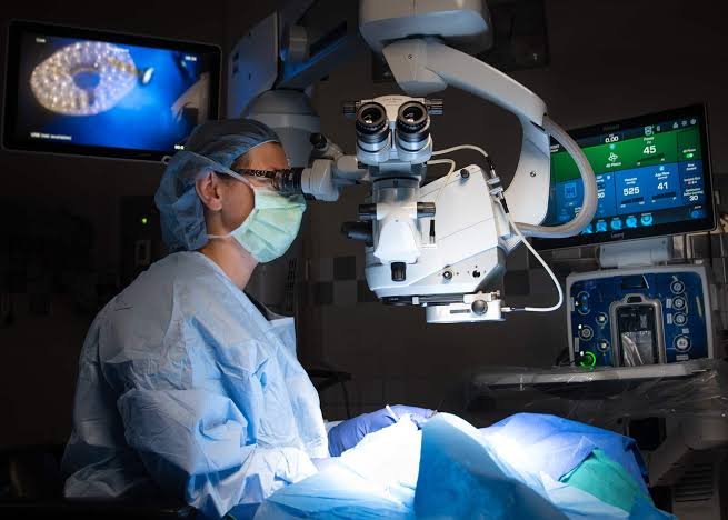
Your approach leverages the eye as a window into neurodegenerative disease. How does retinal imaging provide insights into brain pathology, and what advantages does this strategy offer compared with traditional neuroimaging or blood-based biomarkers?
The retina offers a unique window into neurodegenerative disease because it is a direct developmental extension of the central nervous system. Like the brain, it contains neurons, glia, and synaptic architecture, but unlike the brain, it can be visualized non-invasively and at high resolution during a routine eye exam.
This creates several important advantages. Compared with conventional neuroimaging, retinal imaging is lower cost, more accessible, and more scalable for repeated assessments over time. Compared with blood-based biomarkers, retinal imaging has the potential to provide spatially localised information directly from central nervous system tissue rather than relying on indirect measurements in the periphery, where signal levels may be low and interpretation can be complicated by non-CNS sources. Together, these features make the retina an attractive platform for earlier detection, longitudinal monitoring, and broader access to biomarker-based assessment.
The new $2.5 million Phase 2 grant from the National Institute on Ageing will support further validation work. What are the primary objectives of this phase, and what milestones would demonstrate that the technology is ready to move toward clinical studies in living patients?
The Phase 2 program is designed to build on prior biomarker validation by more fully characterising the three-dimensional distribution of TDP-43-related retinal signals and determining whether those patterns differ across diseases such as ALS, FTD, and LATE. A central goal is to define where these signals are located in the retina, how consistently they can be detected, and whether they show disease-specific spatial signatures that could support diagnosis and patient stratification.
Key milestones would include generating robust 3D retinal maps of TDP-43-associated signals, demonstrating reproducible disease-linked patterns across tissue samples, and developing an AI-enabled analysis platform that can reliably detect and quantify those signals. Together, these advances would establish the biological and analytical foundation needed to design clinical studies in living patients.
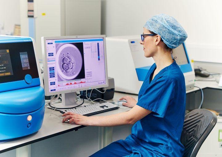
Artificial intelligence will be used to analyse retinal tissue data and identify disease-specific biomarker patterns. How will AI help distinguish between conditions such as Amyotrophic Lateral Sclerosis, Frontotemporal Dementia, and LATE, and what implications could this have for precision medicine?
AI will be used to analyse complex retinal imaging datasets and identify patterns that may be difficult to detect or quantify consistently by human review alone. By integrating signal intensity, spatial distribution, layer-specific localisation, and other image-derived features, AI may help define disease-specific retinal signatures associated with ALS, FTD, and LATE.
If successful, this approach could support a more precise biomarker-based framework for neurodegenerative disease classification. That could improve early identification, help stratify patients for clinical trials, and eventually support more individualised care by linking retinal biomarker patterns with diagnosis, prognosis, or treatment response.
If successfully translated into clinical practice, how might this non-invasive eye test change the diagnostic pathway for patients with suspected neurodegenerative disease, particularly in terms of earlier diagnosis and clinical trial enrollment?
If successfully translated into clinical practice, a retinal biomarker test could meaningfully shorten and strengthen the diagnostic pathway for patients with suspected neurodegenerative disease. Today, many patients undergo prolonged evaluations involving multiple visits, referrals, and exclusion of alternative causes before a diagnosis becomes clear. A non-invasive retinal test could provide earlier biological evidence of disease, helping clinicians identify the right patients sooner and potentially intervene earlier as disease-modifying therapies emerge.
Because retinal imaging is simple, rapid, and already widely available in eye care settings, this approach could also expand access to biomarker-informed evaluation beyond specialised academic centres. Over time, it may also support longitudinal monitoring, patient stratification, and use as an exploratory endpoint in investigational drug development. For clinical trials, better identification of biomarker-positive patients could improve cohort selection, reduce heterogeneity, and increase the ability to detect therapeutic effects.
Looking ahead, do you see the ocular tracer platform expanding beyond TDP-43 detection to target other neurodegenerative biomarkers, and what therapeutic or diagnostic opportunities could this open in the future?
Yes. We view TDP-43 as part of a broader platform strategy focused on mapping molecular biomarkers in the eye that reflect diseases of the brain, eye, and heart. In addition to TDP-43, Amydis is advancing programs targeting amyloid beta and is also exploring tracers for other important proteinopathies, including alpha-synuclein, transthyretin, and phosphorylated tau.
This platform approach could open important diagnostic and therapeutic opportunities. On the diagnostic side, it could enable earlier, more accessible, and more biologically specific detection of multiple diseases. On the therapeutic side, it could support patient selection, treatment monitoring, and biomarker-driven clinical trial design. More broadly, it positions the eye as a practical site for precision medicine approaches across several major protein misfolding disorders.