Pictor Labs is advancing virtual staining technologies that aim to support more integrated tissue analysis workflows, especially as molecular testing and histological assessment continue to compete for limited tissue samples.
In many current workflows, laboratories need to balance the use of tissue for both pathology review and molecular profiling. Conventional chemical staining consumes tissue sections, while molecular testing often requires the same limited specimen for sequencing, spatial transcriptomics, proteomics and other downstream assays. This creates a challenge for laboratories, as every tissue section used for staining may reduce the material available for further testing.
Another key issue is spatial misalignment. In traditional workflows, pathologists may annotate one stained slide, while the molecular assay is performed on an adjacent section. Although the sections are close, they are not exactly the same piece of tissue. This can affect region-of-interest selection and may increase the risk of including tissue areas that are less suitable for testing.
Pictor Labs’ virtual staining approach uses deep learning image translation to generate virtual stained whole-slide images from brightfield or fluorescent scans. The technology is designed to provide stain-specific image views without additional chemical staining, allowing researchers and pathologists to review tissue morphology while preserving more material for downstream molecular workflows.
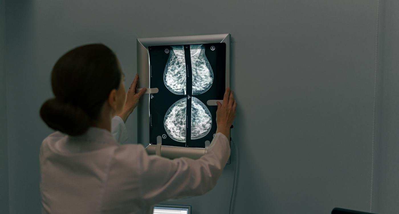
The company’s ClearStain platform focuses on generating virtual H&E images from unstained brightfield slides. This allows assessment and annotation to be performed on the exact section that is going for sequencing, rather than an adjacent approximation. By doing so, the workflow supports better alignment between what the pathologist sees and what is eventually used for molecular analysis.
This is especially important in oncology research and molecular diagnostics workflows, where accurate tissue selection can affect downstream results. Viable tumour areas can support better DNA yield and panel sensitivity, while necrosis, fibrosis and inflammation may reduce sample quality, dilute tumour purity or increase the risk of failed assays. ClearStain aims to help users identify these areas before tissue is extracted for testing.
Pictor Labs also presented internal validation examples showing strong agreement between virtual H&E and traditional histochemical H&E in selected cancer cases. According to the company, internal pilot studies reviewed by board-certified pathologists showed more than 99 percent agreement in tumour region detection and invasive margin identification.
The company’s ReStain platform takes a different approach by generating virtual special stains and IHC-style views from existing H&E slides. This allows researchers to create additional stain perspectives from a single tissue section without new sectioning or repeat wet lab staining. This could be useful for retrospective studies, limited tissue samples, biorepository reuse and multi-step research workflows.
For biopharma research, molecular diagnostic laboratories and digital pathology research, virtual staining highlights a wider opportunity to build more tissue-preserving workflows. It can support faster preliminary review, smarter stain ordering and better use of limited biopsy material, especially as multi-omic research requires more information from smaller tissue samples.
The broader shift is that histological review and molecular extraction may no longer need to compete for the same tissue. With virtual staining, laboratories can gain multiple tissue views from the same sample while preserving more material for molecular analysis. This could be crucial as precision medicine, digital pathology and AI-driven tissue analysis continue to grow.
As the healthcare and research sectors move towards more data-rich and tissue-efficient workflows, platforms such as ClearStain and ReStain reflect the growing role of AI in pathology research. However, the company notes that the technologies discussed are currently for research use only and are not cleared or approved by the FDA for diagnostic use.