
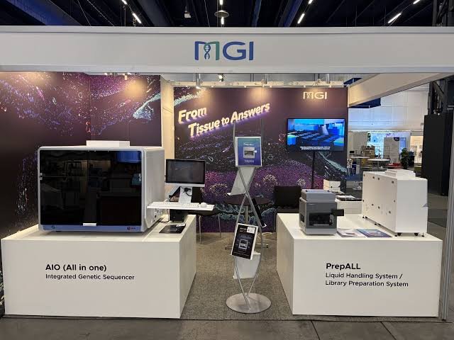
MGI Tech (MGI), a company committed to developing core tools and technologies that drive innovation in life science, announced the European launch of VisiOmics (PMIF-20RS), a fully automated multiplex immunofluorescence (mIF) staining and imaging system, at the 38th European Congress of Pathology (ECP 2026), held in Stockholm from 12 to 16 September 2026.
Under the theme "From Tissue to Answer", the launch anchors a comprehensive pathology portfolio spanning tissue and molecular pathology. Whether the question is asked of proteins in a tissue section or of nucleic acids in a sample, MGI's platforms deliver an accurate answer with AI embedded at every step.
VisiOmics is a fully automated mIF staining and imaging system that brings high-plex, automated tissue analysis within reach of routine laboratory workflows. VisiOmics enables multiplex detection of protein markers within a single tissue section, allowing researchers to characterize cell populations and investigate their spatial organization within the tissue microenvironment:
More than 20-plex detection per run, expandable to more than 100 markers from a single tissue section via cyclic staining
Walk-away automation of the full staining-to-imaging workflow
AI-powered image analysis with FluoXpert Vision — including cell segmentation, phenotyping and spatial analysis — supported by a dedicated AI assistant that lowers the barrier for downstream analysis and data visualization.
"To gain deeper insights into disease biology, pathology research is increasingly combining morphological assessment with multiplex spatial proteomics," said Fang Chen, Vice President of MGI and General Manager of Europe & Africa at MGI. "With VisiOmics now available in Europe, alongside PrepALL and AIO for molecular pathology, we are offering a complete 'From Tissue to Answer' portfolio, whether the starting point is proteins in tissue or nucleic acids in a sample."
MGI is also partnering with the Fraunhofer Institute for Integrated Circuits IIS to demonstrate the compatibility of multiplex immunofluorescence images generated by VisiOmics with MIKAIA®, Fraunhofer IIS's image analysis software for digital pathology and spatial biology research. The collaboration aims to show how researchers can use VisiOmics image data within MIKAIA workflows for cell segmentation, phenotyping and spatial analysis. This work supports greater flexibility in downstream analysis, helping laboratories connect automated tissue imaging with tools suited to their research questions.